Home
Research
Ophthalmic Lab Sites

Ophthalmic Lab Sites

The Shiley Eye Institute is the only academic institution in the San Diego area with comprehensive programs for the clinical care of patients with eye disorders, cutting edge research on surgical techniques and treatments of eye diseases.

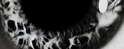
Black and white cornea
Afshari Lab

CORNEAL & OCULAR SURFACE RESEARCH

Advancing research in corneal regeneration, stem cell biology, oxidative stress, genetics, cataract prevention, and dry eye disease.

Visit Afshari Lab
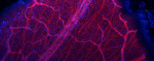
Microscopic image showing a network of red and pink blood vessels on a dark blue background, highlighting fine vascular structures.
Ayyagari Lab

Retinal Molecular Genetics

Finding causative or elusive genes involved in the retinal pathologies and identify therapeutic targets.

Visit Ayyagari Lab

Blurred vertical columns of yellow numbers on a blue gradient background, resembling digital data streams.
Baxter Lab

ophthalmology INFORMATICS

Innovative and cross-disciplinary research in ophthalmology and informatics to develop and leverage advances in health information technology for improving vision and health.
Visit Baxter Lab
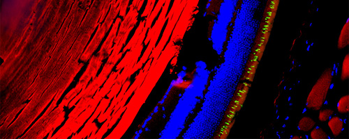
Microscopic image showing a red-stained tissue section on the left with adjacent blue-stained cells and a thin green fluorescent layer, highlighting cellular structures under fluorescence microscopy.
Borooah Lab

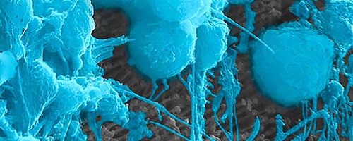
RETINA & MACULA RESEARCH

Leveraging new insights from disease mechanism studies to develop new treatments to prevent blindness caused by these currently untreatable retinal disease.
Visit Borooah Lab
Nanotech Retina
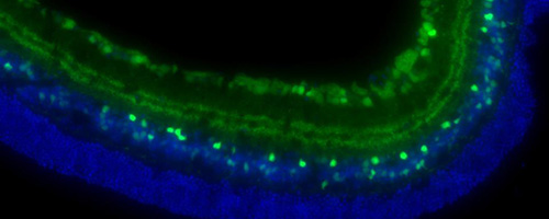
Jacobs Retina Center

NEW TREATMENTS FOR RETINA DISEASE

Seeking solutions for people of all ages who suffer from retina problems, including debilitating disorders such as macular degeneration, diabetic retinopathy, tumors, and inherited diseases.
Visit Jacobs Retina Center
Microscopic image of a curved tissue section showing green fluorescent cells on the outer layer and blue fluorescent cells beneath them.
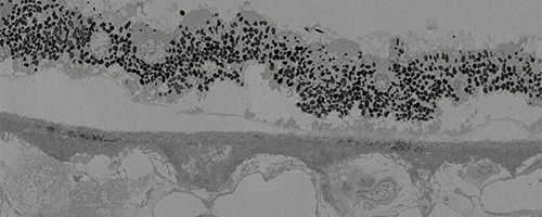
Shaw Lab

Elucidating molecular AMD

Our goal to elucidate the molecular interactions underlying AMD pathogenesis, thereby identifying potential therapeutic targets and possibly establishing potential treatments.
Visit Shaw Lab
Black and white microscopic image showing a densely packed layer of small, dark, rounded cellular structures along a lighter background with varying textures and shapes beneath it.
Toomey Lab

Pharmacological Potential of Glycosaminoglycans

Our lab is focused on understanding the role of Bruch’s membrane glycosaminoglycan changes with aging and the development of early and intermediate AMD.

Visit Toomey Lab
Abstract image of a modern building with overlapping architectural elements and a split color overlay of muted orange and purple tones.
P30 Vision Research Core

Core Grant  //  P30EY022589

NEI funded P30 Vision Research Center Core Grant provides critical support to the UCSD vision research community. The four cores provide access to biostatistical, computational, and technical expertise, as well as sophisticated imaging and tissue processing equipment.
Visit Vision Research
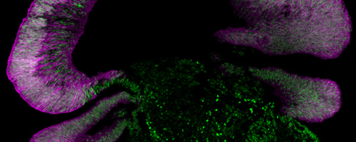
Microscopic image showing purple-stained layered tissue structures surrounding a central dark area with scattered bright green fluorescent spots, suggesting biological or cellular components under fluorescence microscopy.
Wahlin Lab

STEM CELLS & RETINAL REGENERATION

Our lab uses human pluripotent stem cells (PSCs) grown as 3D “mini-retinas” or as dissociated 2D monolayers to investigate this biology.

Visit Wahlin Lab
Profile of a boy looking at a glowing human eye overlaid with a DNA helix and a neural network pattern, against a sunset sky over the ocean.
Treatment Center for Rare Genetic Eye Disease

No Patient Too Rare

Building a new pathway from genetic diagnosis to personalized treatment. We now have tools to identify disease-causing mutations, measure retinal disease with extraordinary precision, and design targeted therapies for ultra-rare genetic conditions.

Visit Rare Genetic Eye Disease Center
Abstract image of a modern building with overlapping architectural elements and a split color overlay of muted orange and purple tones.
Rudell Lab

Translational Pediatric Ophthalmology & Extraocular Muscle Research

Basic science laboratory that studies extraocular muscles, developmental eye disease, and strabismus.

Visit Rudell Lab
Microscopic image showing purple-stained layered tissue structures surrounding a central dark area with scattered bright green fluorescent spots, suggesting biological or cellular components under fluorescence microscopy.
Girkin Lab

RETINA

Lorem ipsum dolor sit amet, consectetur adipiscing elit, sed do eiusmod tempor incididunt ut labore et dolore magna aliqua.

Visit Girkin Lab